ISSN Number
ISSN 2771-019X-

-

Impact Factor
1.2*
ISSN Number
ISSN 2771-019X
Impact Factor
1.2*Doctor of Veterinary Medicine, MVSc, Damot Sore District Agricultural Office, Department of Livestock, Wolaita, Ethiopia.
Doctor of Veterinary Medicine, MVSc, Damot Sore District Agricultural Office, Department of Livestock, Wolaita, Ethiopia.
Email: ejiguhizikel@gmail.com
Received : April 12, 2025,
Accepted : May 05, 2025
Published : May 12, 2025,
Archived : www.jclinmedcasereports.com
Blackleg is an acute bacterial disease affecting ruminants, manifests as a swollen, warm, and painful wound primarily affecting the hind limb. This study presents a case of blackleg in a local breed bull and its treatment outcome. The bull exhibited symptoms such as reduced feed intake, shivering, and lameness, with the onset of the condition reported by the owner in the morning prior to examination. Physical assessment revealed elevated rectal body temperature (40.8°C) and heart rate (100 beats/min), along with lacrimation, lameness, muscle tremors, and congested conjunctival mucous membranes. Treatment involved the administration of procaine penicillin G (20,000 IU/kg/day) for five consecutive days and gentamicin (10 ml) for three consecutive days via intramuscular injection. The bull exhibited a favorable response to the prescribed therapy, ultimately achieving a successful cure upon completion of the treatment regimen. Indeed, the vaccination is frequently used but evidence on the efficacy of vaccination against C. chauvoei to prevent diseases and lethality in cattle is disputed as scant to moderate.
Keywords: Bull; Blackleg; Symptomatic; Damot Sore.
Copy right Statement: Content published in the journal follows Creative Commons Attribution License (http://creativecommons.org/licenses/by/4.0). © Hizikel E (2025).
Journal: The Journal of Clinical and Medical Images, Case Reports (JCMICR) is a fantastic resource for keeping up with the latest clinical advancements and for publishing case reports and clinical images related to a variety of medical illnesses.
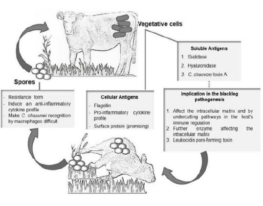
Blackleg is a fatal form of myonecrosis caused by anaerobic, highly pathogenic, endospore- forming, gram-positive bacterium Clostridium chauvoei. It is usually observed in young ruminants and responsible for significant loss in livestock production globally [6,16,20]. Although blackleg vaccination has been carried out in endemic countries, sporadic outbreaks are still recorded annually worldwide. The disease is commonly affecting young flourishing cattle from three months to two years of age. Latent spore of Clostridium chauvoei is thought to be activated through traumatic injury but more localized clostridial myositis can be iatrogenic. C. chauvoei is assumed to be soil borne, but likely does not grow in soil. The bacteria grow readily in the intestinal tract of cattle, and may be recycled through fecal contamination of the soil. Most cases of blackleg occur during the warm months, or after soil excavation, or during very high annual rainfall that can expose and activate latentpores. In addition, the disease is enzootic in areas with a history of flooding [12,20].
The severity of blackleg is ignited by the toxin produced by the bacterium including an oxygen stable haemolysin, an oxygen-labile haemolysin, a DNase (ß-toxin), a hyaluronidase (previously called y-toxin), and a neuraminidase [16,21]. The incubation period of the disease is between 1–5 days [20]. The clinical forms are per acute, which is so short-lived and usually not observed because of sudden death [18]. Acute form is commonly manifested by edema of the heavy muscles and crepitation, lameness and fever (410C) [1,20]. Other unusual findings such as fibrinouspleuritis, pericarditis, epicarditis [2], and severe acute necrotizing enteritis [9] as well as the highly uncommon meningoencephalitis [14,23] are also reported.
Diagnosis of these infections can be made based on clinical picture and postmortem findings. “In Gram stain, all isolates showed numerous short, thick, straight, round-ended, gram-positive rod occurs singly or in short chains” [13]. Anaerobic culture of sections of affected muscle or fluid from the lesion may be employed to identify the causative organism. The bacterium, however, is quite fastidious in its growth requirements. Also, affected tissue is often rapidly overgrown with other clostridial contaminants from the gastrointestinal tract further complicating culture of the causative organism [1]. The use of direct Polymerase Chain Reaction (PCR) was proposed by [4] using common filter paper as an alternative to collecting, storing, and shipping material to the laboratory for the diagnosis of blackleg. Penicillin and gentamicin is the drug of choice in the treatment of black leg in cattle [13]. Annual vaccination of cattle between 6 months and 2 years of age is advocated in areas where the disease is enzootic, just prior to the anticipated danger period.
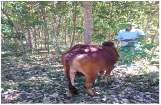
A local breed bull from Sore Mashido kebele was presented to the Gununo veterinary clinic in Wolaita Sodo on February 20, 2023. The owner reported a sudden onset of anorexia (reduced feed intake) and shivering, beginning that morning. The bull was subsequently unable to ambulate (walk).
Clinical examinations and findings
Physical examination revealed a rectal temperature of 40.8°C (hyperthermia) and a heart rate of 100 beats per minute (tachycardia). Bilateral lacrimation (tearing), lameness (claudication), muscle tremors, and congested conjunctival mucous membranes were observed. The animal exhibited good body condition, and other findings were unremarkable.
Diagnosis
Based on the history and clinical findings, the black leg was established tentatively although other acute cases were suspected.
Differential diagnosis
Anthrax, Malignant edema and Heart water from which blackleg was diagnosed tentatively.
Case management and treatment
The bull received intramuscular injections of procaine penicillin G (20,000 IU) daily for five days and gentamicin (10 ml) daily for three days. On the second day of treatment, rectal temperature decreased to 39.2°C and heart rate to 80 bpm, although appetite remained poor. By the third day, both temperature (38.2°C) and heart rate (72 bpm) normalized. Complete clinical recovery was observed upon completion of the treatment regimen (Figure 1).
Blackleg is endemic in Ethiopia and cause overwhelming economic loss in small holder farmers which is associatedwith death, losses in production and reduction in working capacity of farm animals. The current case was asserted as black leg tentatively based on the history and clinical findings (symptomatic) which is similar with symptoms specified in literatures [2,14,20,23]. Accordingly, marked lameness with pronounced swelling of the affected limbs, marked depression, anorexia, ruminalstasis, high pulse rate (100–120/min), high temperature (41OC), marked respiratory distress, emphysema and crepitation of affected heavy muscles are usual in acute form of black leg.
Although many speculative studies have investigated the soluble antigens, particularly toxins, play a significant role in the pathogenesis of blackleg. Currently, five C. chauvoei toxins have been identified, including the hemolytic leukocidin CctA, oxygen-labile hemolysin D (also known as hemolysin III), DNase (referred to as â-toxin), hyaluronidase Nag (formerly known as ã-toxin), and neuraminidase/sialidase NanA. These toxins contribute to the virulence and disease progression of blackleg, exerting various cytotoxic and enzymatic effects that undermine host immunity and tissue integrity.
The pore-forming, oxygen-stable leukocidin hemolysin called C. chauvoei cytotoxin A (CctA) confers strong hemolytic activity, which is observed as a halo around the colonies on blood agar growth medium [5]. CctA as a mature protein has a molecular mass of 32.2 kDa. It is a major toxin and hemolysin produced by C. chauvoei, which is shown to be highly cytotoxic to the bovine epithelial cell line ECaNEp [5]. The antibodies directed against CctA play the main role in the protective immunity exerted against blackleg; thus, it is a valuable candidate for blackleg vaccines and for the potency testing of current vaccines.
The previously described oxygen-stable necrotizing hemolysin (α-toxin) (Hang’ombe et al., 2006) might be CctA, although the reported molecular mass of this α-toxin hemolysin is 25kDa, which is significantly lower. Alternatively, this α-toxin could represent the putative hemolysin III, also called hemolysin D or δ-toxin (protein #276) found on the genome of C. chauvoei [6], whose molecular mass is around 25 kDa. However, the latter might correspond to a weak hemolysin that is oxygen labile and potentially thiol-activated [7]. It must be noted that hemolysin III is not specific to C. chauvoei as several pathogenic, commensal, and environmental gram-positive bacteria express it or carry genes coding for this class of hemolysin. Although there is no clarity about hemolysin III in C. chauvoei, it is reported to be similar to the θ-toxin produced by C. perfringens and the tetanolysin produced by C. tetani [10]. Using monospecific antibodies directed against CctA, Frey et al., [5] fully neutralized all the hemolytic activity expressed by C. chauvoei, showing that other than CctA, this pathogen does not produce any entity with measurable hemolytic activity.
DNase (ß-toxin) and hyaluronidase (called y-toxin) toxins are greatly responsible for the pathogenesis of the disease. DNase (ß-toxin) is an enzyme of the deoxyribonuclease and responsible for the nuclear degradation of muscle cells and actively participates in clostridialmyonecrosis. While, hyaluronidase toxin is assumed to be responsible for the destruction of the loose connective tissue that surrounds the muscles, thus favoring the spread of C. chauvoei in the tissues of the infected and the end products of hyaluronate degradation are disaccharides, which might be a source of nutrients for the pathogen [23].
Neuraminidase/sialidase (NanA) was purified and characterized by Heuermann et al., [11]. They showed that the enzymatic activity cleaves N-acetylneuraminic acid (sialic acid) in carbohydrate polymers, present in many mammalian cell membranes as well as many microorganisms [19]. NanA was characterized in detail as an 81-kDa protein that is secreted as a dimer [22]. It is encoded by the nanA gene, which is fully conserved across the C. chauvoei strains isolated over 60 years from various geographical locations across four different continents [17]. A recombinant molecule derived from nanA containing the sialic acid-binding domain (CBM40) is able to fully neutralize the sialidase activity of C. chauvoei [22]. Thus, NanA can also be used as a potential antigen to aid protective immunity.
A case of blackleg was successfully treated with a combination of procaine penicillin G (20,000 IU intramuscularly for five consecutive days) and gentamicin (10 ml intramuscularly for three days). Significant clinical improvement was observed within 48 hours, consistent with previous case reports [3,15].
Blackleg is a disease that predominantly affects young, rapidly growing cattle, often resulting in sudden and significant livestock losses. While blackleg can manifest in cattle as young as 2 months old, the highest incidence of cases is observed in cattle aged between 6 months and 2 years. Although humans are generally not susceptible to this disease, caution is advised when handling deceased or suspected blackleg-infected animals due to potential risks. The economic impact of blackleg can be severe in various regions globally. Indeed, the vaccination is frequently used but evidence on the efficacy of vaccination against C. chauvoei to prevent diseases and lethality in cattle is disputed as scant to moderate.
Based on the above conclusion the following recommendations are forwarded:
• Animals must be vaccinated against blackleg to avoid loss.
• Veterinarians should promptly diagnose cases clinically or through laboratory testing and administer appropriate treatment.
• During outbreaks, community members should immediately report suspected cases to veterinary authorities, who must then differentiate blackleg from other diseases before implementing vaccination programs.
• Governmental support should include adequate vaccine and drug distribution, along with training for veterinary personnel.
• Public education should promote safe carcass disposal (incineration or deep burial without opening) to prevent further transmission.